Remember that moment when you truly appreciated your eyesight? For me, it was a recent scare, a fleeting blur after too many hours glued to a screen, that hammered home the irreplaceable value of clear vision.
It also deepened my fascination with the incredible leaps we’re seeing in ophthalmic diagnostic equipment. We’re standing on the cusp of an eye care revolution, where cutting-edge technology isn’t just improving diagnoses, but fundamentally changing how we prevent and manage eye diseases.
From the almost magical precision of AI-powered retinal scanners that can spot a problem years before symptoms appear, to sophisticated telehealth platforms making expert eye care accessible in remote areas, the landscape is electrifying.
As someone who’s personally witnessed these innovations unfold, what truly strikes me is how these advancements move beyond simple detection. They empower us with unprecedented insights, leading to highly personalized treatment plans and dramatically improved patient outcomes, turning potential blindness into preserved sight.
These aren’t just tools; they’re guardians of our future vision. Let’s explore this landscape accurately below.
The Silent Guardians: How Early Detection is Redefining Eye Health

The sheer power of early detection in ophthalmology is something I find profoundly compelling. It’s not just about catching a problem; it’s about preventing a crisis.
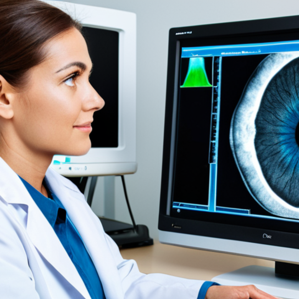
When I first saw an Optical Coherence Tomography (OCT) scan revealing the earliest, almost imperceptible changes in a patient’s retina, I was genuinely awestruck.
This wasn’t merely a picture; it was a three-dimensional map of incredibly delicate tissue, revealing secrets years before a traditional eye chart ever would.
This kind of technology empowers eye care professionals to intervene with remarkable precision, often before any noticeable vision loss occurs. It feels like we’re finally getting ahead of the curve, instead of constantly playing catch-up with progressive diseases.
My personal brush with vision blurriness made me realize how much we take our sight for granted, and seeing these machines in action makes me breathe a sigh of relief, knowing that so many people can avoid the scarier parts of that journey.
It’s an investment in a future where preventable blindness becomes a relic of the past, driven by tools that see what the naked eye cannot.
1. Unveiling the Microscopic: Optical Coherence Tomography (OCT)
OCT is a game-changer, plain and simple. It uses light waves to capture cross-sectional images of the retina, optic nerve head, and anterior segment. Think of it as a non-invasive biopsy without the biopsy part.
For conditions like glaucoma, macular degeneration, and diabetic retinopathy, OCT provides unparalleled detail. I recall a colleague showing me an OCT scan of a patient who felt perfectly fine, yet the scan revealed subtle fluid buildup beneath the macula – a tell-tale sign of early wet AMD.
Without that OCT, the patient might have lost significant central vision before even realizing something was wrong. This level of insight allows for immediate intervention, preserving precious sight that would otherwise be irreversibly damaged.
It’s truly a marvel how a light beam can render such intricate details of the eye’s delicate structures.
2. The AI Revolution in Retinal Imaging
If OCT provides the map, AI is the seasoned guide pointing out the hidden trails. AI-powered diagnostic tools are transforming how we analyze retinal images, making detection faster, more accurate, and accessible.
These algorithms can identify patterns indicative of various eye diseases, often with greater consistency than human interpretation, especially in high-volume screenings.
I’ve witnessed systems that can flag diabetic retinopathy with astounding precision from a simple fundus photo, and not just in a clinic – some are designed for primary care settings, bridging gaps in rural areas.
It’s not about replacing the human expert but augmenting their capabilities, freeing them to focus on complex cases and patient care. This synergy between human expertise and artificial intelligence is creating an unprecedented safety net for global eye health, making diagnostic capabilities available in places where specialized ophthalmologists might be scarce.
Beyond the Walls: The Rise of Tele-Ophthalmology and Remote Diagnostics
The idea of expert eye care reaching you, rather than you having to reach it, was once a distant dream for many. But now, it’s a tangible reality, especially in the vast landscapes of North America where specialized clinics can be hours away for some communities.
Tele-ophthalmology isn’t just a convenience; it’s a lifeline, expanding access to critical diagnostic services far beyond traditional clinic walls. My personal experience with telehealth during the pandemic, even for routine check-ups, made me deeply appreciate the power of technology to connect us.
When it comes to eye care, this connection means early detection and continuous monitoring for individuals who might otherwise fall through the cracks due to geographical barriers or limited mobility.
It’s truly democratizing eye health, ensuring that a professional assessment isn’t a luxury, but an accessible right.
1. Bridging Gaps with Remote Retinal Screening
Imagine a mobile unit, equipped with a digital retinal camera, visiting community centers or remote primary care clinics. Patients get their eyes photographed on-site, and those images are then securely transmitted to an ophthalmologist hundreds of miles away for review.
This isn’t science fiction; it’s happening right now, dramatically improving screening rates for conditions like diabetic retinopathy, which can silently steal sight.
I’ve heard countless stories from colleagues about patients in underserved areas who, thanks to these programs, received a timely diagnosis of a treatable condition that would have otherwise progressed unchecked.
This model reduces the burden of travel, saves time and money for patients, and most importantly, saves sight. It’s a pragmatic solution that leverages technology to overcome real-world hurdles.
2. Virtual Consultations and Continuous Monitoring
Tele-ophthalmology also extends to virtual consultations for follow-up appointments or for discussing initial screening results. Secure video conferencing platforms allow patients to speak directly with an eye care professional, show them how they’re feeling, and receive personalized advice.
For chronic conditions, remote monitoring devices, such as at-home visual field tests or tonometers for glaucoma, are becoming more prevalent. This allows for more frequent data collection, providing a richer, real-time picture of a patient’s condition between in-person visits.
It truly gives patients a sense of empowerment and control over their health journey, fostering a partnership with their eye care team that goes beyond episodic clinic visits.
Tailoring Treatment: The Precision of Personalized Eye Care Diagnostics
The days of one-size-fits-all medical approaches are quickly fading, and nowhere is this more evident than in modern ophthalmology. My own journey, which involved a brief but intense period of anxiety about my vision, made me acutely aware of how much better it feels when care is truly tailored to *you*.
Today’s advanced diagnostic equipment doesn’t just identify a problem; it provides such a nuanced understanding of an individual’s unique ocular anatomy and pathology that treatments can be customized with incredible precision.
This isn’t just about better outcomes; it’s about a more efficient and less invasive path to preserving vision, minimizing side effects, and truly optimizing the patient’s experience.
It feels like a genuine evolution from reactive treatment to proactive, personalized care.
1. Corneal Topography and Wavefront Analysis: Precision for Refractive Surgery
For anyone considering vision correction procedures like LASIK or PRK, detailed diagnostics of the cornea are paramount. Corneal topography creates a detailed map of the corneal surface, identifying irregularities that could impact surgical outcomes.
Wavefront analysis, on the other hand, measures how light travels through the eye, detecting even subtle aberrations that traditional prescriptions might miss.
I once met someone who had undergone a refractive surgery that corrected not just their basic prescription but also an obscure night vision issue, all thanks to the granular data provided by these sophisticated machines.
This level of detail allows surgeons to plan custom-tailored procedures that maximize visual acuity and minimize post-operative complications, leading to genuinely stunning results and a new level of clarity.
2. Genetic Testing and Biomarker Discovery in Ocular Diseases
Beyond structural imaging, the frontier of personalized eye care is pushing into genetics and molecular diagnostics. Identifying specific genetic markers or biomarkers in a patient can predict disease progression, determine responsiveness to certain therapies, and even reveal predisposition to conditions like age-related macular degeneration or certain types of glaucoma.
This insight is transformative. It means treatments can be initiated proactively for high-risk individuals, or specific, highly targeted therapies can be chosen, avoiding a trial-and-error approach.
It feels like we are on the verge of decoding the eye’s most intricate secrets, unlocking pathways to truly preventive and highly effective interventions.
The Core Pillars: Essential Diagnostic Tools in Every Eye Clinic
While the flashiest new technologies often grab the headlines, it’s important to remember the foundational instruments that form the bedrock of daily eye care.
These tried-and-true devices, often enhanced with modern digital capabilities, are indispensable for comprehensive examinations and are used countless times a day in clinics around the world.
As someone who’s spent time observing optometrists and ophthalmologists at work, I’ve seen firsthand how these tools, from the seemingly simple to the deceptively complex, work in concert to build a complete picture of a patient’s ocular health.
They might not always be “futuristic,” but their consistent reliability and diagnostic power are absolutely critical for maintaining vision for millions.
1. Slit Lamp Biomicroscopy: The Indispensable Workhorse
The slit lamp is arguably the most fundamental and versatile piece of equipment in an eye clinic. It allows the eye care professional to examine the anterior segment (eyelids, conjunctiva, cornea, iris, lens) and, with the aid of special lenses, the posterior segment (retina, optic nerve).
It’s essentially a high-magnification microscope with a powerful light source that can be manipulated into a thin “slit” of light, allowing for a three-dimensional view of ocular structures.
I remember vividly how an optometrist, using just a slit lamp, spotted a tiny, almost invisible foreign body on my friend’s cornea after a windy day, preventing a much larger issue.
It’s the first stop for almost every eye complaint and its diagnostic utility is simply unmatched for a comprehensive, hands-on examination.
2. Tonometry: Measuring the Silent Threat of Glaucoma
Intraocular pressure (IOP) measurement, or tonometry, is crucial for detecting and managing glaucoma, a leading cause of irreversible blindness. There are various methods, from the gold standard Goldmann applanation tonometry, which gently touches the eye, to non-contact tonometry (the “air puff” test familiar to many).
While the air puff can be a little startling, its quick, non-invasive nature makes it ideal for screening. Elevated IOP doesn’t automatically mean glaucoma, but it’s a significant risk factor, and consistent monitoring is key.
The simplicity of the test belies its profound importance in identifying a condition that progresses without symptoms until significant vision is lost.
It’s a testament to how seemingly simple tests can be powerful guardians of our sight.
| Diagnostic Tool Category | Key Function | Benefits for Patients |
|---|---|---|
| Advanced Imaging (OCT, Fundus Camera) | Detailed structural and photographic analysis of the retina and optic nerve. | Early detection of diseases like AMD, glaucoma, diabetic retinopathy, often before symptoms appear. Non-invasive and quick. |
| Refractive Diagnostics (Topography, Wavefront) | Precise mapping of corneal shape and optical aberrations. | Optimized outcomes for refractive surgeries (LASIK, PRK), improved visual quality, especially for complex cases. |
| Intraocular Pressure Measurement (Tonometry) | Measures the pressure inside the eye. | Crucial for screening and monitoring glaucoma, which often has no early symptoms. Prevents irreversible vision loss. |
| Tele-Ophthalmology Platforms | Remote eye examinations and consultations. | Increased access to care for individuals in remote areas, reduced travel burden, convenient follow-ups. |
Empowering Patients: Understanding Your Diagnostic Journey
It’s easy to feel a bit overwhelmed when you’re in an eye clinic, with all the different machines and tests. I know I did, especially during that moment of vision uncertainty.
But here’s the thing: understanding what each diagnostic tool does and why it’s being used isn’t just about curiosity; it’s about active participation in your own health.
When patients are empowered with knowledge, they become partners in their care, asking better questions, adhering to treatment plans, and feeling more confident about their journey.
This isn’t just a professional talking; it’s deeply rooted in my personal belief that informed patients are healthier patients.
1. Preparing for Your Eye Exam: What to Expect from Diagnostics
Knowing what to expect can significantly reduce anxiety during an eye exam. Typically, a comprehensive exam involves several steps. After initial vision acuity tests, you might experience the “air puff” for tonometry.
Then comes the slit lamp examination, where the doctor looks closely at your eye. Depending on your age, risk factors, or symptoms, you might have pupils dilated for a better view of the retina, followed by an OCT scan or a fundus photograph.
It’s a series of non-invasive, quick procedures designed to build a complete picture of your eye health. If you have questions, never hesitate to ask your eye care professional to explain what each machine does.
They’re usually more than happy to clarify.
2. The Role of Patient Data in Personalized Treatment Plans
The data collected from these diagnostic tests isn’t just a jumble of numbers and images; it’s the foundation upon which your personalized treatment plan is built.
For example, if your OCT shows signs of fluid buildup, your doctor might recommend specific eye drops or injections. If tonometry reveals consistently high pressure, they might prescribe glaucoma medication.
This data allows for highly specific and effective interventions, tailored precisely to your unique condition. I’ve heard countless stories where detailed diagnostic information helped avert a serious vision crisis, all because the doctors had the right data to make informed decisions.
It highlights how integral these tools are to achieving truly effective, individualized patient care.
Future Horizons: What’s Next in Ophthalmic Innovation
The pace of innovation in ophthalmic diagnostics is nothing short of breathtaking. What was once considered futuristic is now standard, and what’s currently in the labs promises to revolutionize eye care even further.
My fascination with this field only deepens when I consider the incredible potential for new technologies to address currently unmet needs, pushing the boundaries of what’s possible in preserving sight.
It’s not just about incremental improvements; it’s about paradigm shifts that will redefine how we approach eye disease, moving towards even more proactive, predictive, and personalized interventions for everyone.
1. Wearable Tech and Continuous Monitoring
Imagine contact lenses embedded with sensors that continuously monitor intraocular pressure and wirelessly transmit data to your eye doctor, or smart glasses that detect subtle changes in your visual field throughout the day.
This isn’t far-fetched. Prototypes and early-stage devices are already in development, promising a future where continuous, real-time monitoring of key eye health parameters becomes commonplace.
This level of data could allow for incredibly early detection of disease progression, enabling interventions even before a patient notices any symptoms, completely transforming the management of chronic eye conditions.
This would truly be a game-changer for conditions that require constant vigilance.
2. Advanced AI and Machine Learning in Predictive Analytics
While AI is already making waves in image analysis, the next frontier lies in predictive analytics. Imagine AI systems that can analyze a patient’s entire medical history, lifestyle factors, genetic profile, and diagnostic test results to predict their individual risk of developing certain eye diseases decades in advance.
Such systems could also forecast the progression of existing conditions with unprecedented accuracy, guiding preventative strategies and treatment timings.
This shift from reactive diagnosis to truly proactive, personalized risk assessment will allow for interventions that are not just early, but optimally timed, providing a new layer of protection against vision loss.
It’s a thrilling prospect for a future where eye care is less about treating disease and more about preventing it.
Concluding Thoughts
The journey through the intricate world of ophthalmic diagnostics truly brings into focus the profound impact of proactive eye care. What began with that initial awe at an OCT scan has only deepened into a conviction that these technologies aren’t just tools; they are vital partners in safeguarding our most precious sense.
Empowering ourselves with knowledge, embracing these innovations, and fostering open communication with our eye care professionals ensures that we’re not just reacting to vision challenges, but actively shaping a future where clear sight is preserved for generations.
It’s a collective effort, and one that gives me immense hope.
Useful Information for Your Eye Health Journey
1. Prioritize Regular Eye Exams: Even if your vision seems fine, comprehensive eye exams are crucial for detecting silent diseases like glaucoma and diabetic retinopathy in their earliest stages. Think of it as a vital health check, not just a vision test.
2. Don’t Ignore Subtle Symptoms: Any persistent vision changes, discomfort, or unusual visual phenomena (like floaters or flashes) warrant a prompt visit to your eye care professional. Early action can make a significant difference in outcomes.
3. Ask Questions About Your Diagnostics: When your doctor recommends a test like an OCT or visual field, don’t hesitate to ask what it measures and why it’s important for your specific case. Understanding empowers you to be an active participant in your care.
4. Embrace a Vision-Healthy Lifestyle: Beyond clinic visits, simple habits like a balanced diet rich in antioxidants, protecting your eyes from UV light, and taking screen breaks can contribute significantly to long-term eye health.
5. Explore Tele-Ophthalmology Options: For follow-ups or initial screenings, especially if you live remotely, inquire about tele-ophthalmology services. They can provide convenient access to expert care without the need for extensive travel.
Key Takeaways
Modern ophthalmic diagnostics, spearheaded by advanced imaging like OCT and the revolutionary integration of AI, are transforming eye care from reactive treatment to proactive prevention.
These powerful tools enable incredibly early detection of diseases, often before symptoms even manifest, significantly improving treatment outcomes and preserving vision.
Furthermore, tele-ophthalmology is democratizing access to expert care, bridging geographical gaps, while personalized diagnostics like corneal topography and genetic testing allow for highly customized and effective interventions.
Ultimately, these innovations empower both eye care professionals and patients, fostering a future where preventable blindness becomes increasingly rare and individualized vision preservation is the norm.
Frequently Asked Questions (FAQ) 📖
Q: The opening paragraph highlighted
A: I-powered retinal scanners that can spot problems “years before symptoms appear.” How exactly are these cutting-edge AI diagnostic tools revolutionizing early detection for common, potentially sight-threatening eye conditions?
A1: From what I’ve personally witnessed and heard from specialists, these AI-powered retinal scanners are nothing short of revolutionary. Think about conditions like diabetic retinopathy or glaucoma, which often progress silently, with damage accumulating long before you notice any blur or vision loss.
It’s terrifying, really. But these AI systems? They can analyze a retinal scan with such incredible detail and speed, picking up on minute changes – tiny hemorrhages, subtle nerve fiber layer thinning – that a human eye, even an expert one, might miss on a routine check, especially if it’s an early stage.
It’s like having an impossibly meticulous second pair of eyes that can process vast amounts of data in seconds. The true game-changer here is that pre-symptomatic detection.
It shifts the entire paradigm from reacting to damage to proactively preserving sight. I’ve spoken to folks who, thanks to these early flags, were able to start treatment for conditions they didn’t even know they had, potentially saving years, even decades, of good vision.
It’s not just an improvement; it’s a profound shift in how we prevent blindness.
Q: The text also mentions sophisticated telehealth platforms making expert eye care accessible in remote areas. How are these technological advancements making specialist eye care more widely available, particularly for those facing geographical or mobility challenges?
A: This one truly resonates with me because I’ve seen the struggles firsthand. For someone living far from a major city, or for an elderly relative with limited mobility, a routine eye exam can be an all-day ordeal – travel, waiting, the exhaustion of it all.
Telehealth platforms are literally shrinking those distances. It’s not just a video call with your doctor; it’s a comprehensive system where initial screenings can be done closer to home, with images and data securely transmitted to an ophthalmologist hundreds of miles away.
They can then review, consult, and even provide preliminary diagnoses without the patient ever having to leave their local clinic or even their home, in some cases with portable devices.
I know of a community in a fairly isolated part of New England, for instance, where access to specialized eye care used to mean a five-hour drive. Now, thanks to these platforms, residents can get expert opinions and follow-ups much more readily.
It’s an absolute game-changer for accessibility, breaking down barriers that previously meant delayed care or no care at all.
Q: You describe these advancements as “guardians of our future vision.” Beyond just early detection and accessibility, what’s the fundamental impact of these technologies on actual patient outcomes and the long-term preservation of sight?
A: This is where the real magic happens, in my opinion. It goes far beyond simply “finding” a problem. The sophisticated data collected by these advanced diagnostic tools – things like optical coherence tomography (OCT) and advanced perimetry – provides an incredibly detailed, almost three-dimensional map of your eye’s health.
This granular insight allows specialists to create truly personalized treatment plans. Instead of a generic approach, doctors can tailor interventions, monitor progression with unprecedented precision, and adjust therapies in real-time.
I’ve heard countless stories where this level of detail has made a colossal difference. For someone with early-stage macular degeneration, for example, knowing the exact rate of cellular change means the difference between timely, less invasive intervention and potentially irreversible vision loss.
It transforms reactive treatment into proactive, highly targeted care. These aren’t just tools; they empower doctors to be precision strategists, turning potential blindness into genuinely preserved, functional sight, giving people a far better chance at maintaining their quality of life for decades to come.
📚 References
Wikipedia Encyclopedia
구글 검색 결과
구글 검색 결과
구글 검색 결과
구글 검색 결과
구글 검색 결과