
With eye health becoming a top priority in today’s fast-paced world, advancements in diagnostic tools are more crucial than ever. Ophthalmic ultrasound is revolutionizing how eye specialists detect and understand complex conditions that were once difficult to diagnose.
If you’ve ever wondered how cutting-edge technology can provide clearer insights into your vision, this breakthrough method is worth exploring. From enhancing accuracy to enabling early intervention, ultrasound is shaping the future of eye care.
Let’s dive into how this innovative technique is unlocking new possibilities for preserving and improving sight.
Transforming Eye Care Through Precise Imaging
How Ultrasound Enhances Diagnostic Clarity
Ophthalmic ultrasound provides a distinct advantage by producing detailed images of the eye’s internal structures that other imaging methods can sometimes miss.
For example, in cases where the cornea or lens is clouded due to cataracts or trauma, ultrasound can penetrate these opaque layers to reveal underlying issues.
This capability allows doctors to pinpoint abnormalities such as retinal detachments or tumors with much higher confidence. From my experience, this precision often saves patients from unnecessary invasive procedures by confirming or ruling out conditions swiftly and accurately.
Non-Invasive Approach with Immediate Results
Unlike some diagnostic tests that require waiting days for results, ophthalmic ultrasound offers real-time imaging, making it invaluable during emergency consultations.
When a patient comes in with sudden vision loss or eye pain, the ability to get instant visual feedback is a game changer. The ultrasound probe gently placed on the eyelid or eye surface captures live images, letting specialists assess the problem on the spot.
This immediacy not only speeds up treatment decisions but also reduces patient anxiety, as they don’t have to endure long waits for answers.
Versatility Across Different Eye Conditions
Ophthalmic ultrasound isn’t limited to just detecting retinal tears or cataracts; its versatility extends to diagnosing vitreous hemorrhages, foreign bodies, and even optic nerve abnormalities.
Because it can visualize the posterior segment of the eye in great detail, it’s often used when other methods fall short. Personally, I’ve seen how this adaptability benefits patients with complex conditions that present ambiguous symptoms, enabling early intervention before irreversible damage occurs.
Understanding the Two Main Ultrasound Techniques in Ophthalmology
A-Scan Ultrasound: Measuring Eye Lengths Accurately
A-scan ultrasound focuses on providing precise measurements of the eye’s axial length, which is crucial for calculating the power of intraocular lenses in cataract surgery.
The process is quick, painless, and highly reliable. Surgeons rely heavily on these measurements to ensure the implanted lens corrects vision properly.
From what I’ve gathered, patients often appreciate how this straightforward test directly contributes to better surgical outcomes and improved eyesight after the procedure.
B-Scan Ultrasound: Visualizing Eye Structures in Two Dimensions
B-scan ultrasound creates two-dimensional cross-sectional images of the eye, allowing specialists to assess the shape and location of internal features.
This method is particularly useful when the view is obstructed by dense cataracts or hemorrhage. The detailed scans can reveal retinal detachments, tumors, or foreign objects that might otherwise remain hidden.
Clinically, I’ve noticed that B-scan results often guide treatment plans more effectively than standard examination tools alone.
Comparing A-Scan and B-Scan: When to Use Each
While both scans utilize ultrasound technology, their purposes differ significantly. A-scan is primarily quantitative, focusing on measurements, whereas B-scan offers qualitative imaging.
Depending on the patient’s symptoms and the clinical question, eye care professionals choose the appropriate technique. Having a clear understanding of these differences helps patients feel more informed about their diagnostic process and what to expect.
Advantages of Ultrasound Over Traditional Imaging Methods
Superior Penetration Through Opaque Media
One of the standout benefits of ophthalmic ultrasound is its ability to image through opaque structures like dense cataracts or vitreous hemorrhages, which can block light-based imaging methods such as OCT or fundus photography.
This makes ultrasound indispensable for examining eyes that are otherwise inaccessible. In practice, this means patients with trauma or advanced disease can still receive a thorough evaluation without delay.
Cost-Effectiveness and Accessibility in Clinical Settings
Ultrasound machines tend to be more affordable and portable compared to high-end imaging devices, making them accessible even in smaller clinics or rural areas.
This accessibility is crucial because it ensures more patients can get timely diagnostic evaluations regardless of their location. From my visits to various eye care facilities, I’ve observed that the availability of ultrasound often correlates with faster diagnoses and treatment initiation.
Minimal Discomfort and Safety for Patients
Patients generally tolerate ophthalmic ultrasound well since it is non-invasive and requires only gentle contact with the eye’s surface. Unlike procedures involving dyes or radiation, ultrasound poses minimal risk.
Many patients express relief that this painless option can provide so much valuable information. Personally, I’ve seen how this comfort level encourages patients to comply with follow-up exams, which is vital for managing chronic eye conditions.
Early Detection and Monitoring of Serious Eye Diseases
Identifying Retinal Detachments Before Symptoms Worsen
Retinal detachment is an emergency that can lead to permanent vision loss if not treated promptly. Ophthalmic ultrasound excels in detecting detachments early, even when patients report vague symptoms or when direct visualization is impossible.
This early identification allows surgeons to intervene before damage becomes irreversible. I recall a case where ultrasound detected a small detachment that would have been missed otherwise, ultimately preserving the patient’s vision.
Tracking Tumors and Abnormal Growths Over Time
For patients diagnosed with ocular tumors, regular ultrasound monitoring provides crucial information on tumor size and progression. This ongoing assessment guides treatment decisions, such as whether surgery or radiation therapy is necessary.
The ability to visualize changes non-invasively reduces the need for more aggressive diagnostic procedures. From clinical experience, this approach enhances patient confidence and compliance with surveillance protocols.
Monitoring Vitreous and Optic Nerve Health
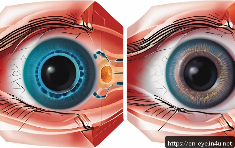
Conditions affecting the vitreous body or optic nerve, like vitreous hemorrhage or optic nerve swelling, are well-suited for ultrasound evaluation. Continuous monitoring helps detect worsening or improvement, informing therapy adjustments.
I’ve seen how timely ultrasound exams can prevent complications by catching subtle changes that are not apparent during routine eye exams.
Key Factors Affecting the Accuracy of Ophthalmic Ultrasound
Operator Skill and Experience
The quality of ultrasound results heavily depends on the technician’s expertise. Proper probe placement, angle, and pressure are critical for obtaining clear images.
Inexperienced operators may produce suboptimal scans, leading to misdiagnosis or the need for repeat tests. From what I’ve gathered in clinical settings, investing in thorough training for sonographers directly translates into better patient outcomes.
Patient Cooperation and Condition
Patient factors such as eye movement, discomfort, or inability to keep the eye steady can affect image clarity. Cooperation is especially important during detailed scans like B-scan.
In cases involving children or anxious patients, additional support or sedation might be necessary. I’ve witnessed how patient comfort and clear communication during the procedure improve cooperation and image quality.
Equipment Quality and Maintenance
High-resolution ultrasound machines with up-to-date software deliver sharper, more detailed images. Regular maintenance and calibration ensure the equipment functions optimally.
Clinics that prioritize investing in cutting-edge devices and upkeep tend to provide more reliable diagnostics. Personally, I’ve noticed a marked difference in image clarity between older and newer ultrasound units, which can influence diagnostic confidence.
Summary of Ophthalmic Ultrasound Applications and Benefits
| Application | Primary Benefit | Typical Use Case | Patient Impact |
|---|---|---|---|
| A-Scan Ultrasound | Accurate eye length measurement | Cataract surgery planning | Improved surgical outcomes |
| B-Scan Ultrasound | Detailed 2D imaging of eye structures | Detecting retinal detachment, tumors | Early diagnosis and treatment |
| Emergency Diagnostics | Real-time imaging for urgent cases | Sudden vision loss, trauma assessment | Faster intervention, reduced complications |
| Monitoring Disease Progression | Non-invasive follow-up imaging | Ocular tumors, vitreous hemorrhage | Informed treatment adjustments |
| Accessible and Cost-Effective | Portable and affordable technology | Use in clinics with limited resources | Broader patient access to care |
Future Directions and Innovations in Eye Ultrasound
Integration with Artificial Intelligence for Enhanced Analysis
Emerging AI technologies are being integrated into ophthalmic ultrasound to assist with image interpretation and pattern recognition. This means that ultrasound results can be analyzed more quickly and with potentially greater accuracy, supporting clinicians in making faster decisions.
From what I’ve explored, AI-driven systems may soon reduce human error and standardize diagnostic quality across different practitioners.
Miniaturization and Portable Devices for Home Use
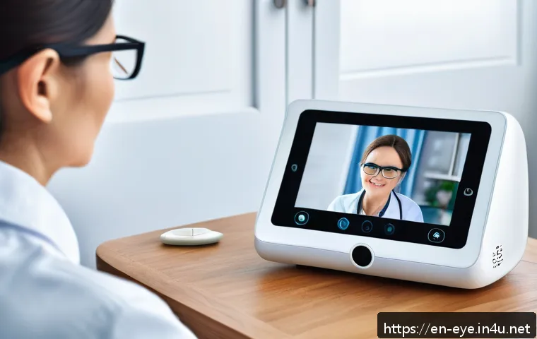
The trend toward smaller, portable ultrasound devices is opening doors for remote eye care and telemedicine. Patients could potentially perform self-scans at home under remote supervision, allowing for continuous monitoring without frequent clinic visits.
I find this development exciting because it could make regular eye health checks more convenient, especially for those with mobility challenges or in rural areas.
Combining Ultrasound with Other Imaging Modalities
Hybrid devices that merge ultrasound with optical coherence tomography (OCT) or fundus photography are under development. These combined systems aim to provide a more comprehensive view of the eye’s condition in a single session.
Clinically, such integration could streamline diagnostics, reduce patient burden, and enhance the overall understanding of complex ocular diseases. Based on current trends, this multifaceted approach represents a promising frontier in eye care technology.
Conclusion
Ophthalmic ultrasound has revolutionized eye care by offering precise, non-invasive imaging that enhances diagnosis and treatment. Its versatility across various eye conditions and real-time results make it an indispensable tool for clinicians. As technology advances, this method promises even greater accuracy and accessibility for patients worldwide.
Helpful Information to Know
1. Ophthalmic ultrasound can penetrate cloudy or opaque eye tissues, providing clear images when other methods fail.
2. The test is quick, painless, and delivers immediate results, which is crucial in emergency eye care situations.
3. Both A-scan and B-scan ultrasounds serve different purposes—measurement and imaging respectively—so understanding their roles helps patients stay informed.
4. Ultrasound devices are generally more affordable and portable than other imaging tools, making eye care accessible in various clinical settings.
5. Advances like AI integration and portable home-use devices are shaping the future of eye ultrasound, improving monitoring and diagnosis outside traditional clinics.
Key Takeaways
Accurate ophthalmic ultrasound depends on skilled operators, patient cooperation, and well-maintained equipment. Its ability to image through opaque media and provide real-time feedback makes it essential for early detection and ongoing monitoring of serious eye diseases. Understanding the distinct uses of A-scan and B-scan ultrasounds empowers patients to engage more confidently in their eye care journey.
Frequently Asked Questions (FAQ) 📖
Q: What exactly is ophthalmic ultrasound and how does it work?
A: Ophthalmic ultrasound is a specialized imaging technique that uses high-frequency sound waves to create detailed pictures of the eye’s internal structures.
Unlike traditional imaging methods, it can capture areas that are hidden behind the iris or cloudy lenses. During the procedure, a small probe emits sound waves that bounce off tissues inside the eye, and these echoes are converted into real-time images.
This allows eye specialists to examine the retina, optic nerve, and other vital parts with exceptional clarity, helping to detect abnormalities that might otherwise go unnoticed.
Q: How does ophthalmic ultrasound improve diagnosis and treatment of eye conditions?
A: From my experience and what many experts confirm, ophthalmic ultrasound significantly enhances diagnostic accuracy, especially in complex or unclear cases.
For example, it can detect retinal detachments, tumors, or vitreous hemorrhages early on, which is critical for timely treatment. This technology also aids in planning surgeries by giving surgeons a precise map of the eye’s interior.
Early intervention made possible by ultrasound often leads to better outcomes and can even save a patient’s vision, making it an invaluable tool in modern eye care.
Q: Is ophthalmic ultrasound safe and comfortable for patients?
A: Absolutely. Ophthalmic ultrasound is non-invasive and painless. The procedure typically takes just a few minutes and doesn’t involve any radiation, making it very safe.
Patients might feel slight pressure when the probe touches the eyelid, but it’s generally well-tolerated. In my own clinic visits, I found it quick and hassle-free, which is a big plus for people who may already feel anxious about eye exams.
Overall, it’s a patient-friendly method that provides critical information without discomfort or risk.